Existing Patients
(908) 850-4200
New Patients
(908) 275-5307


At the office of Paulussen Dental, we use modern diagnostic tools to deliver confident, predictable care. One of the cornerstones of our imaging capabilities is cone-beam computed tomography (CBCT), a three-dimensional X-ray technology that reveals details conventional radiographs cannot. By integrating CBCT into our diagnostic workflow, we improve treatment planning and reduce uncertainty for many common and complex dental procedures.
CBCT is not a replacement for routine X-rays, but a powerful supplement when depth, spatial relationships, and an accurate view of anatomy matter. The technology produces high-resolution volumetric images of teeth, jawbones, sinuses, and surrounding structures in a single, efficient scan. That clarity helps our team make better-informed decisions while maintaining a focus on safety and patient comfort.
Cone-beam computed tomography captures a cone-shaped beam of X-rays as the scanner rotates around the patient’s head. The resulting data are reconstructed into a volumetric image — essentially a stack of thin slices — which can be viewed in three orthogonal planes or as a 3D model. This makes it possible to see the exact shape and position of teeth, roots, and bone with precise spatial context.
The value of CBCT lies in its ability to reveal relationships that are difficult or impossible to judge on two-dimensional films. For example, the proximity of a tooth root to the inferior alveolar nerve, the width and height of available bone for an implant, or the presence of impacted teeth and their angulation are all seen more clearly. That level of detail reduces guesswork and supports targeted, minimally invasive care.
Clinically, CBCT images are used as a roadmap. They allow clinicians to measure distances, evaluate bone density patterns, and identify anatomical variations before any treatment is started. This visual precision translates into fewer surprises during procedures and more predictable outcomes for patients.
CBCT is particularly helpful when routine exams and standard X-rays don’t provide enough information to form a confident diagnosis or treatment plan. Common indications include implant planning, complex extractions of impacted teeth, assessment of jaw pathology, and evaluation of the airways and sinuses. It’s also used in endodontics to locate additional canals, identify root fractures, or assess periapical lesions.
Orthodontic cases that require an understanding of skeletal relationships and airway volume can also benefit from CBCT. Likewise, patients with a history of trauma, suspected bone lesions, or atypical anatomy often require the enhanced visualization CBCT offers. Because the scan is localized, clinicians can target the region of interest to minimize exposure while getting the information needed.
Ultimately, the decision to recommend CBCT is based on clinical necessity. Our goal is to order imaging that directly informs diagnosis or guides treatment decisions — not imaging for its own sake. When the information from a CBCT scan will change the approach to care, the scan becomes an essential part of the diagnostic process.
CBCT also plays a role in interdisciplinary planning. When restorative, surgical, or orthodontic specialists collaborate, a shared 3D data set improves communication and coordination. The result is a comprehensive plan that accounts for function, esthetics, and long-term stability.
For dental implants, CBCT is a standard of care in many practices because it shows available bone volume, vital structures, and angulation for optimal implant placement. With this information, clinicians can determine the best implant size and orientation and identify any need for bone grafting or sinus lift procedures before surgery.
In oral surgery, CBCT helps map the exact location of impacted teeth, cysts, or bony irregularities. Surgeons can plan incisions and bone removal with greater confidence, reducing operative time and preserving healthy tissues when possible. The three-dimensional view also assists in avoiding critical anatomy and anticipating potential complications.
When restorative or prosthetic work requires precise occlusion and tooth positioning, CBCT data can be integrated with digital impressions and CAD/CAM workflows. This coordination allows for restorations that fit better and function more predictably, particularly in full-arch reconstructions or complex bite rehabilitations.
Any imaging that uses X-rays involves a trade-off between diagnostic benefit and radiation exposure. Modern CBCT units are engineered to minimize dose by allowing region-specific scans and by using protocols optimized for dental applications. The radiation from a dental CBCT scan is generally higher than a single periapical film but significantly lower than medical CT scans.
Before recommending a CBCT scan, our clinicians assess whether the diagnostic advantage outweighs the exposure. For many patients, the scan replaces multiple conventional films and provides richer information with a single, quick acquisition. Protective measures and contemporary low-dose settings further reduce exposure while preserving image quality.
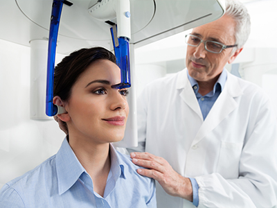
We also take steps to make the experience comfortable. Scans are brief — often under a minute — and the patient remains seated or standing with gentle support for the head. Clear instructions and the attentive presence of our team help ensure a smooth, calm process for patients of all ages.
CBCT images are a tool, not a standalone diagnosis. Interpreting the data requires clinical correlation with the patient’s history, examination findings, and other imaging. Our clinicians review the scans with a focus on actionable findings: issues that will influence treatment choice, timing, or sequence.
When a consultative read by a radiology specialist is needed, we collaborate with qualified imaging experts to ensure comprehensive evaluation. This multidisciplinary approach is particularly useful for complex pathology, suspected lesions that require biopsy, or cases where surgical risks must be fully mapped out.
We make it a point to review significant findings with patients in plain language, showing relevant slices or 3D views when helpful. Visual explanations help patients understand the reason for a recommended treatment and how the planned approach addresses the problem. That transparency supports informed decision-making and builds confidence in the care plan.
Our team at Paulussen Dental integrates CBCT thoughtfully into patient care, using the images to reduce uncertainty and improve outcomes. When combined with clinical expertise and contemporary technologies, CBCT helps us deliver safer, more precise dentistry.
In summary, cone-beam computed tomography is a powerful diagnostic asset that enhances planning and execution for a range of dental treatments. If you have questions about whether CBCT is appropriate for your situation or would like to learn more about how we use this technology in our practice, please contact us for more information.
CBCT stands for cone-beam computed tomography, a three-dimensional imaging technique that captures volumetric X-ray data of the teeth, jaws, sinuses and surrounding structures. Unlike standard periapical or panoramic films, CBCT creates a stack of thin slices that can be viewed in multiple planes or reconstructed into a 3D model for precise spatial context. That capability reveals relationships and anatomic details that two-dimensional images cannot reliably show.
The enhanced visualization helps clinicians evaluate root positions, bone quantity and nearby vital anatomy with greater confidence. Because the data are volumetric, measurements such as bone height, width and angulation can be made directly from the images. This makes CBCT a powerful supplement to routine imaging when depth and three-dimensional relationships matter for diagnosis or treatment planning.
CBCT is recommended when conventional X-rays do not provide sufficient information to make a confident diagnosis or plan treatment. Typical indications include implant planning, assessment of impacted or atypically positioned teeth, evaluation of jaw pathology, and complex endodontic cases where root anatomy or fractures are suspected. Clinicians also use CBCT for airway analysis, trauma assessment and interdisciplinary treatment planning.
The decision to order CBCT is based on clinical necessity rather than routine use, with the goal of obtaining information that will directly influence care. Because scans can be localized, the imaging field is restricted to the area of interest to minimize exposure. When the additional information will change the treatment approach, CBCT becomes an essential diagnostic tool.
For implant treatment, CBCT provides a clear picture of available bone volume, bone morphology and the location of critical structures such as the inferior alveolar nerve and sinus cavities. This allows clinicians to select appropriate implant length, diameter and angulation before surgery, reducing the risk of intraoperative surprises. The ability to visualize cross-sectional anatomy helps identify whether additional procedures like bone grafting or a sinus lift will be required.
CBCT datasets can also be integrated with digital impressions and surgical planning software to fabricate guided surgical guides or provisional restorations. This digital workflow improves precision during placement and supports predictable prosthetic outcomes. By planning in three dimensions, clinicians can minimize invasiveness and improve long-term implant success.
As with any X-ray-based modality, CBCT involves ionizing radiation, so the decision to image balances diagnostic benefit against exposure. Modern dental CBCT units and protocols are optimized to limit dose by allowing region-specific scans and reducing scan times, and their exposure is generally higher than a single periapical film but substantially lower than a medical CT scan. Clinicians follow the principle of justification and optimization—ordering scans only when clinically indicated and using the lowest reasonable dose for the required diagnostic quality.
Protective practices include selecting a smaller field of view whenever possible, adjusting settings for patient size and using contemporary low-dose protocols. Pregnant patients and other sensitive populations are assessed individually to determine whether imaging is necessary or if alternatives exist. Open communication with the clinical team ensures patients understand why a scan is recommended and how exposure is managed.
A CBCT appointment is typically quick and noninvasive, with scans often completed in less than a minute from the time the unit begins rotating. The patient remains seated or standing while the scanner rotates around the head, and gentle supports or positioning aids are used to minimize movement. Clear instructions are provided before and during the scan to help achieve a motion-free acquisition and optimal image quality.
There is no direct contact, no intraoral sensors, and most patients experience little or no discomfort beyond keeping still for a short period. After the scan, images are reconstructed and reviewed by the treating clinician, who will explain any significant findings and how the images will inform diagnosis or treatment planning. At Paulussen Dental the team emphasizes patient comfort and clear communication throughout the process.
CBCT interpretation is performed by dentists with appropriate training in three-dimensional imaging as part of their clinical workflow, and when complex pathology is suspected a specialist radiologist or oral and maxillofacial radiologist may be consulted. Accurate interpretation requires correlating image findings with the patient’s history, clinical examination and other diagnostic data to form a complete clinical picture. Clinicians trained in implantology, endodontics or oral surgery commonly use CBCT as an advanced diagnostic aid within their scope of practice.
When a consultative read is needed, clinicians collaborate with qualified imaging experts to ensure a thorough evaluation of lesions, anatomic variations or surgical risks. This multidisciplinary approach helps clarify ambiguous findings and guides safe treatment planning. Patients are informed of significant results in plain language and are shown relevant slices or 3D reconstructions when helpful for understanding the proposed care.
Yes. CBCT can be a valuable adjunct in endodontics because it provides three-dimensional views of root canal anatomy, canal curvatures and periapical tissues that are often obscured on two-dimensional films. The technology can reveal extra canals, complex canal morphologies, vertical root fractures and the extent of periapical lesions with greater clarity. This information helps clinicians decide whether nonsurgical retreatment, surgical intervention or alternative therapies are indicated.
Because small fields of view and high-resolution protocols are available for endodontic applications, clinicians can tailor imaging to capture fine anatomic details while limiting exposure. Interpretation always occurs in the context of clinical signs and symptoms to ensure that radiographic findings are meaningful for patient care. When CBCT alters the working diagnosis or treatment strategy, it serves as a decisive diagnostic resource.
In orthodontics, CBCT provides a complete view of skeletal relationships, tooth positions and temporomandibular joint anatomy, which can inform treatment planning for complex cases. Three-dimensional scans help evaluate impacted teeth, asymmetries and the spatial relationships between the maxilla and mandible, supporting more precise mechanics and staging. CBCT is particularly useful when traditional records do not explain the underlying skeletal or dental issue.
CBCT can also contribute to airway assessment by visualizing the nasal passages, nasopharynx and pharyngeal airspace. While it should not replace comprehensive sleep medicine evaluation, CBCT data can help identify anatomic contributors to airway restriction and support referrals for further testing when appropriate. As always, CBCT findings are integrated with clinical evaluation and specialist input to determine their significance for treatment.
No. CBCT is a complementary tool rather than a replacement for conventional dental imaging. Two-dimensional radiographs such as bitewings and periapical films remain the first-line modality for routine screening, caries detection and many standard diagnostic tasks because they are fast, low-dose and clinically sufficient for those purposes. CBCT is reserved for situations where three-dimensional information will materially affect diagnosis or treatment planning.
Appropriate imaging selection depends on the question being asked; clinicians choose the simplest exam that answers that question reliably. By using CBCT selectively and in conjunction with other imaging types, dental teams provide efficient, evidence-based care while minimizing unnecessary exposure. This balanced approach ensures patients receive the right image at the right time for their individual needs.
CBCT improves treatment outcomes by reducing uncertainty and enabling more precise, individualized treatment plans. The detailed visualization of bone, roots and anatomic landmarks helps clinicians anticipate challenges, select optimal surgical approaches and avoid vital structures, which can decrease operative time and complications. When combined with digital planning tools, CBCT data support guided surgery and prosthetic workflows that enhance accuracy and restorative fit.
Beyond procedural planning, CBCT facilitates interdisciplinary communication by providing a shared three-dimensional dataset for restorative, surgical and orthodontic teams. That coordination leads to comprehensive plans that balance function, esthetics and long-term stability. When used thoughtfully, CBCT is an integral part of delivering predictable, high-quality dental care.