Existing Patients
(908) 850-4200
New Patients
(908) 275-5307


Digital radiography has transformed how dentists see and treat oral health. At Paulussen Dental, we use digital X-ray systems that combine high-resolution sensors with secure software to capture, view, and store diagnostic images in moments — improving patient comfort and clinical accuracy without adding complexity to your visit.
Digital radiography captures the same diagnostic information as traditional film X-rays but does so through electronic sensors and computer processing. Those sensors convert X-ray energy into a digital file that can be enhanced, measured, and archived. The result is a clearer picture of tooth structure, root anatomy, bone levels, and areas of decay that are not visible during a visual exam alone.
Because images are digital, clinicians can zoom, adjust contrast, and annotate in real time to highlight areas of concern. These tools help dentists evaluate subtle changes more reliably and explain findings to patients using on-screen visuals. For routine checkups, restorative planning, or monitoring healing after treatment, digital images make it easier to spot problems early and track progress over time.
Beyond immediate diagnostics, digital images integrate into an electronic health record so each image becomes part of a patient’s longitudinal chart. That continuity supports more informed decision-making and makes consultations with specialists more productive when referral or collaboration is needed.
One of the most meaningful advantages of digital radiography is the reduction in radiation exposure compared with conventional film techniques. Digital sensors are more sensitive to X-rays, so they require less radiation to generate a diagnostic-quality image. This improvement makes routine imaging safer for both children and adults while still delivering the detail clinicians need.
Safety goes beyond radiation numbers. Digital workflows eliminate chemical developers and paper-based handling, reducing environmental hazards in the operatory. The streamlined process also minimizes the time a patient spends holding sensors in place, which can improve comfort, especially for those with sensitive gag reflexes or limited patience.
Dental teams follow established guidelines for imaging frequency and protective practices, ensuring each exposure is clinically justified. In other words, digital radiography helps us balance the need for clear diagnostic information with a commitment to patient safety and conservative use of imaging.
With digital systems, images appear on-screen almost instantly after exposure. That rapid turnaround shortens appointments when imaging is needed and allows dentists to review findings with patients while the information is still fresh. Instead of waiting for film development, care decisions can be discussed on the same visit.
Digital files are easy to share securely with labs, specialists, or other providers when collaborative care is required. Whether coordinating an implant restoration, consulting on periodontal treatment, or sending images for an orthodontic evaluation, the ability to transfer high-quality images quickly improves coordination and reduces delays in treatment planning.
Patients also benefit from seeing their own images. Pointing out an early cavity, a hairline fracture, or the margin of an old filling on-screen helps people understand the rationale for proposed treatment and participate actively in care decisions.
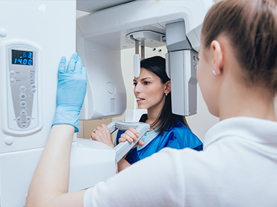
Digital radiography typically relies on either intraoral sensors or phosphor plate systems. An intraoral sensor resembles a small electronic pad that rests inside the mouth; when exposed, it transmits data instantly to the computer. Some practices use wireless sensors that improve patient comfort and simplify positioning. In every case, image capture is followed by immediate digital processing to optimize clarity and diagnostic value.
Once acquired, images are stored within a secure practice management system or imaging cloud that maintains patient privacy and supports backups. Modern dental software includes safeguards such as user permissions and encrypted transmission to protect sensitive health information. This infrastructure makes it straightforward to retrieve past films during follow-up care or to compare current and previous images side by side.
Routine maintenance and calibration of sensors and software ensure consistent image quality. Dental teams follow manufacturer guidance and regulatory standards to keep equipment functioning properly — because high-quality images depend on both good technique and reliable technology.
Digital radiography supports a more patient-centered experience in several practical ways. Faster imaging means less time in the chair, and clearer images help limit unnecessary repeat exposures. For anxious patients, efficient workflows and clear visual explanations can reduce uncertainty and improve cooperation during examinations.
The technology also expands what can be offered during a typical appointment. For example, immediate imaging can assist with same-day restorations or targeted treatment planning when swift clinical decisions are needed. From pediatric patients to adults managing complex restorative needs, digital radiography adapts to a wide range of clinical scenarios while keeping comfort and quality at the forefront.
Finally, the eco-friendly nature of going digital — no chemical development and less physical storage — aligns with a modern approach to care that values both clinical excellence and environmental responsibility.
In summary, digital radiography is a safe, efficient, and precise tool that enhances diagnosis, communication, and overall patient care. Our office leverages these capabilities to improve clinical outcomes and make visits smoother and more informative. For more information about how we use digital imaging or to discuss whether it is right for your needs, please contact us to learn more.
Digital radiography refers to dental X-ray systems that capture images with electronic sensors and convert them into digital files for immediate viewing and analysis. These images reveal tooth structure, root anatomy, bone levels, and areas of decay that are not visible during a visual exam alone. Clinicians use digital images for routine checkups, restorative planning, surgical preparation, and monitoring healing over time.
Because the images are electronic, they can be enhanced, measured, and archived in a patient record to support long-term care. Digital radiography also supports patient communication by displaying findings on-screen so patients can see the same visuals their clinician uses. The approach streamlines diagnosis and helps guide more informed treatment decisions.
Digital radiography captures the same diagnostic information as film-based X-rays but uses sensors and computer processing instead of photographic film and chemical development. The sensors convert X-ray energy into digital data that can be adjusted for contrast, magnified, and annotated, offering clearer and more flexible images for diagnosis. This immediate digital output eliminates the wait time and physical storage associated with film.
Because sensors are more sensitive to X-rays than film, digital systems typically require less radiation to produce a diagnostic image while maintaining image quality. Digital files are also easier to share with specialists or dental labs, improving collaboration and reducing delays in care coordination. Overall, the workflow is faster, more adaptable, and more environmentally friendly than film processing.
Yes. Digital sensors are more sensitive to X-rays than traditional film, which allows clinicians to use lower exposure settings while still obtaining diagnostic-quality images. Dental teams follow established guidelines and protective practices to ensure each exposure is clinically justified and minimized whenever possible.
The reduced radiation dose, combined with modern positioning techniques and protective shielding, makes routine imaging safe for most patients, including children and adults. If you have specific concerns about radiation exposure, your clinician can explain the benefits and precautions taken for your individual situation.
Digital dental images are captured using intraoral sensors or phosphor plate systems positioned in the mouth or with extraoral sensors for panoramic or cone beam imaging. Intraoral sensors resemble small electronic pads that transmit data instantly to a computer; some systems are wireless to improve comfort and positioning. After exposure, the image is processed by software that optimizes clarity, adjusts contrast, and prepares the file for review.
Once processed, images are stored in the practice management system or in a secure imaging cloud where they can be compared side by side with prior studies. Routine maintenance and calibration of sensors and software ensure consistent image quality and reliable interpretation. Good technique, combined with properly maintained equipment, produces the high-quality images clinicians rely on for diagnosis.
Digital radiography enhances diagnosis by producing clearer, manipulable images that reveal subtle changes in tooth and bone structure that can be missed on a visual exam. Tools such as zoom, contrast adjustment, and measurement features help clinicians detect early decay, hairline fractures, bone loss, and root anatomy with greater precision. These capabilities support more accurate diagnoses and earlier intervention when needed.
For treatment planning, immediate access to high-quality images shortens the decision-making process and facilitates same-visit discussions about options and next steps. Digital files can be shared with specialists or dental labs to coordinate complex care such as implant planning, endodontic treatment, or orthodontic referrals. The result is a more collaborative and efficient planning process that benefits patient outcomes.
Digital images are stored within secure practice management systems or encrypted imaging clouds that include user permissions, secure backups, and protected transmission protocols. These technical safeguards help maintain patient privacy and reduce the risk of unauthorized access. Staff access is typically restricted to authorized users, and routine audits help ensure security practices remain current.
In addition to technical protections, dental teams follow policies for record retention, secure sharing, and HIPAA-compliant communication when transferring images to specialists or labs. Patients may request copies of their images, and clinicians can provide explanations while maintaining confidentiality. Together, these measures safeguard personal health information throughout the imaging lifecycle.
Yes. Digital systems produce images almost instantly after exposure, eliminating the delay associated with film development and allowing clinicians to review findings during the same appointment. This faster turnaround can shorten visits when imaging is required and support on-the-spot decisions such as same-day restorations or prompt treatment adjustments.
Fewer repeat exposures are needed because digital images can be optimized immediately, and improved positioning options—such as wireless sensors—can increase patient comfort and cooperation. The combined efficiency of capture, processing, and review helps make appointments more productive and less time-consuming for patients.
Yes. Common systems include intraoral digital sensors for bitewing and periapical images, phosphor plate systems that use reusable plates scanned after exposure, and extraoral options such as panoramic and cone beam computed tomography (CBCT) that provide broader views of the jaws. Each system serves specific diagnostic needs, from detecting cavities to evaluating bone volume for implants or assessing complex anatomy.
The choice of system depends on the clinical question, patient comfort, and the level of detail required for treatment planning. Clinicians select the appropriate imaging modality to balance diagnostic value with patient safety, and many practices maintain multiple systems to address a wide range of cases.
Imaging frequency is individualized based on a patient’s oral health status, risk factors, age, and clinical findings rather than a fixed schedule. Preventive patients with low risk may require radiographs less often, while those with active disease, restorative needs, or orthodontic concerns may need more frequent monitoring. Dental teams follow professional guidelines to determine when imaging is clinically justified.
During routine exams, your clinician will review your history and current condition and recommend imaging only when it will influence diagnosis or treatment. This tailored approach seeks to minimize exposure while ensuring clinicians have the information needed to provide safe, effective care.
Paulussen Dental integrates digital images into the patient record so files can be securely shared with specialists, labs, or consultants to support collaborative treatment planning. High-quality, transferable images allow outside providers to review anatomy and pathology in detail, which helps streamline referrals for implant placement, endodontic treatment, or periodontal care. Quick electronic transfer reduces delays and supports more cohesive interdisciplinary management.
When collaboration is needed, the practice prepares images with appropriate annotations and clinical notes to clarify findings and treatment goals. This coordinated approach improves communication between providers and helps patients move through complex care pathways with clearer expectations and consistent records.