Existing Patients
(908) 850-4200
New Patients
(908) 275-5307


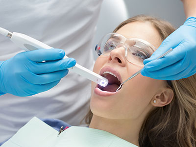
An intraoral camera is a compact, pen-sized imaging device designed to capture detailed, full-color pictures of the teeth, gums, and other soft tissues inside the mouth. Unlike a simple mirror, the camera provides high-resolution, close-up views that reveal subtle surface features—tiny cracks, early enamel breakdown, worn restorations, and areas of concern that can be difficult to spot with the naked eye alone. Images are displayed in real time on a monitor so both clinician and patient can view the same vantage point.
Because the camera can magnify structures many times their actual size, it exposes details that improve diagnostic accuracy. The device uses integrated lighting and precision optics to produce consistent images even in tight spaces. These live images support a more thorough oral exam by complementing tactile assessment and digital radiography, giving the care team a fuller picture of oral health.
Beyond simply “seeing,” intraoral cameras turn visual observations into recordable data. Single-frame captures and short video sequences can be saved to a patient’s chart, creating a visual timeline that documents changes over time. This capability makes the intraoral camera a practical tool for monitoring healing, tracking disease progression, and communicating results clearly with patients and other members of the treatment team.
During a routine exam or when investigating a specific concern, the clinician will gently guide the camera around the mouth to capture the problem areas. The procedure is quick and noninvasive: the camera’s small size allows it to reach behind molars and along the gumline to record angles that are otherwise difficult to inspect. Patients can watch images appear on the screen as they are taken, which helps demystify clinical findings and fosters a collaborative conversation about options and next steps.
As the care team reviews images together with the patient, they can annotate or zoom in on particular features to explain why a finding matters. For example, a dark spot on a tooth may be magnified to show surface breakdown, or a fringe of inflamed tissue can be highlighted to demonstrate early gum disease. This shared visual reference helps patients understand the rationale for recommended care and allows clinicians to document their clinical observations precisely.
The captured images become part of the permanent record and are available for second opinions or specialist consultations when needed. If a restorative procedure is planned—such as a crown, onlay, or implant restoration—images help the laboratory and specialists coordinate color, shape, and fit more effectively. The saved photos also support continuity of care by providing future clinicians with an accurate baseline to compare against later visits.
One of the primary advantages of intraoral imaging is improved diagnostic confidence. The magnified, well-lit views make it easier to detect small fractures, early caries, failing margins around restorations, and subtle soft-tissue changes. Catching problems earlier often results in less invasive treatment and better long-term outcomes for patients.
For treatment planning, photographs serve as a precise visual reference that complements X-rays and three-dimensional scans. Combining multiple imaging modalities creates a more complete clinical picture, allowing clinicians to devise treatment plans that are tailored to the tooth anatomy and the patient’s overall oral condition. This integrated approach reduces guesswork and helps set realistic expectations.
From a patient education perspective, seeing a magnified image of their own mouth is frequently more persuasive than a verbal description alone. Visuals make it easier to understand why care is being recommended and how specific treatments will address the issue. This transparency encourages informed decision-making and often improves patient adherence to preventive measures and prescribed therapies.
Additionally, visual documentation is useful for monitoring changes over time. By comparing images taken at different appointments, clinicians can assess the progression or resolution of lesions, the stability of restorations, and the effectiveness of periodontal therapy, enabling timely intervention when adjustments are needed.
Modern intraoral cameras combine high-quality optics, LED illumination, and digital sensors to produce crisp, color-accurate images with minimal distortion. Many systems offer adjustable magnification, autofocus, and built-in white-balance controls so the clinician can capture consistent images regardless of lighting conditions. These technological refinements translate into dependable visuals that are clinically useful and easy for patients to interpret.
Image files are stored within secure dental software platforms and integrated with electronic health records for streamlined access. Compatibility with digital radiography, CBCT, and CAD/CAM systems enables clinicians to cross-reference images and plan restorative work with precision. The result is a more efficient workflow and a coordinated approach to complex cases.
In terms of safety, intraoral cameras are designed to meet strict infection-control standards. The outer sheaths or sleeves used during exams are single-use or sterilizable according to manufacturer protocols, protecting both patients and staff. The process of capturing images is painless and noninvasive, making intraoral imaging suitable for patients of all ages—including those who may be anxious about more intrusive diagnostic tools.
Patients will typically notice the camera’s small tip as the clinician moves it briefly around the mouth; the experience is quick and comfortable. There is no special preparation required before the exam, and images can be captured at routine checkups, during focused problem assessments, or as part of pre- and post-procedure documentation. Because the camera captures live views, clinicians can immediately point out and explain areas of concern.
Those images remain accessible in the patient’s record for future reference, which helps when tracking outcomes or coordinating care with specialists. For example, if restorative work is planned, intraoral photos can be shared with a dental laboratory or a referring specialist to ensure that prosthetics match the patient’s anatomy and esthetic goals. Visual documentation also helps the team evaluate the success of treatments over time.
Finally, using intraoral images supports a more transparent and informed care process. Patients leave the appointment with a clearer understanding of their oral health status and the reasons behind any recommended treatment. If you have questions about how intraoral imaging will be used during your visit, our team can explain the process and review any images with you in detail so you feel confident about your care.
At Paulussen Dental, we incorporate intraoral imaging into comprehensive exams to enhance communication, improve diagnostic clarity, and support precise treatment planning for patients in Hackettstown and the surrounding communities. If you’d like to learn more about how this technology may be used during your next appointment, please contact us for more information.
An intraoral camera is a small, pen-sized imaging device that captures high-resolution, full-color images of teeth, gums and other soft tissues inside the mouth. It uses LED illumination and precision optics to produce clear, magnified views that reveal surface details smaller than the naked eye can see. Images are displayed in real time on a monitor so clinicians and patients can review findings together.
The camera records single frames or short video sequences with digital sensors that integrate into dental practice software. Clinicians can adjust magnification and white balance to maintain consistent, color-accurate photos even in tight spaces. Saved images become part of the patient record and create a visual timeline for monitoring change over time.
Magnified, well-lit images from an intraoral camera make it easier to detect small fractures, early caries and marginal defects around restorations. Seeing these features at high resolution increases diagnostic confidence and often enables treatment at an earlier, less invasive stage. The camera complements tactile exams and radiographs by revealing surface characteristics that X-rays do not show.
Clear photographs also help clinicians document findings precisely and communicate their clinical significance to patients. Annotating or zooming in on areas of concern aids treatment planning by clarifying the extent and location of a problem. This combined visual and clinical documentation supports more targeted, efficient care.
Intraoral imaging is considered safe and noninvasive because it captures visible light images rather than using ionizing radiation. The procedure is painless and quick, making it suitable for routine checkups as well as focused assessments. Modern devices meet medical device safety standards for electronics and optics.
Infection control protocols further protect patients: disposable sheaths or sterilizable components are used according to manufacturer guidelines. Images are managed within secure dental software platforms to protect privacy and confidentiality. If you have specific concerns about device safety or sterilization, the clinical team can explain their protocols before imaging.
During the exam, the clinician will gently guide the camera around your mouth to capture the necessary views, typically taking only a few minutes. You may watch the images appear on a monitor as they are captured, which helps you understand what the clinician is observing. No special preparation is required and the experience is generally comfortable for patients of all ages.
The clinician may pause to zoom or annotate an image to explain a finding and discuss treatment options in real time. Captured images are saved to your record for future comparison or for sharing with specialists when appropriate. This immediate visual feedback supports clearer, more informed conversations about care.
Intraoral images are stored within secure dental practice software that integrates with electronic health records and complies with applicable privacy regulations. Access is limited to authorized clinical staff and protected by the practice's recordkeeping and cybersecurity policies. Images are saved as part of the patient chart to create an accurate clinical history.
When images are shared for referrals or laboratory communication, they are transmitted through secure channels or included in the official patient record to maintain confidentiality. Clinics typically retain images according to medical record retention policies to support continuity of care. You may request copies of your images as part of your record access rights.
Yes. High-quality intraoral photos are a practical tool for planning restorative procedures because they capture surface texture, shade and soft-tissue relationships that matter for esthetics and fit. These visuals help dental laboratories match color and shape and assist specialists in assessing the clinical situation before treatment. When combined with radiographs and digital scans, intraoral images contribute to precise, coordinated planning.
Photographs also provide a baseline record to evaluate the fit and condition of restorations over time, making it easier to spot changes that may require intervention. For complex cases, images can be included with digital workflows to inform CAD/CAM design and verify outcomes. This integration reduces guesswork and improves predictability.
Yes. One of the main benefits of intraoral imaging is that patients can view the same images as the clinician during the appointment, which promotes transparency and education. Being able to see a magnified view of a problem area often makes explanations clearer than verbal descriptions alone. This shared view encourages questions and helps patients make informed decisions about their care.
The clinician can highlight specific features, annotate images and compare them with previous photos to illustrate progression or healing. Discussing images in real time allows you to understand the rationale for recommended care and to agree on next steps. If you want further clarification, the team can save annotated images in your record for later review.
Intraoral cameras are well suited for children and patients who experience dental anxiety because the procedure is noninvasive and quick, with no discomfort. The live images can be used as a gentle educational tool to show young patients what is happening and to build trust through age-appropriate explanations. Clinicians can adjust their technique to minimize movement and make the experience comfortable.
For anxious patients, being able to see and understand findings often reduces uncertainty and helps demystify clinical recommendations. Photography can also document progress in behavior-based treatment plans, providing measurable milestones that support a calm, structured approach to care. The clinical team can combine imaging with other comfort measures to support a positive visit.
Intraoral photography and radiographic imaging serve complementary roles in diagnosis and treatment planning: photographs reveal surface detail and color while X-rays and CBCT scans show internal structures and bone. Using both modalities gives clinicians a fuller picture of tooth integrity, root anatomy and surrounding bone, which improves decision-making. This multimodal approach reduces the likelihood of missing clinically relevant information.
Images can be cross-referenced with radiographs and three-dimensional scans within integrated dental software to plan restorations or surgical procedures with greater precision. For example, photos help translate radiographic findings into a visual context for shade selection, margin assessment and soft-tissue management. Together, these tools support safer, more predictable outcomes.
At Paulussen Dental in Hackettstown, intraoral imaging is incorporated into comprehensive exams and used routinely to document findings and educate patients about their oral health. Clinicians rely on these images to clarify diagnoses, coordinate care with laboratories and specialists, and to monitor treatment outcomes over time. The practice integrates intraoral photos with digital radiography and charting to create a cohesive clinical record.
If you would like intraoral images taken during your next visit, the team will explain the process and review the pictures with you so you understand any recommendations. Saved images become part of your permanent record and can be used for follow-up comparisons or specialist consultations as needed. Contact the office if you have questions about how intraoral imaging will be used during your care.